Category Archive Newsletter

Deep Vein Thrombosis

Deep Vein Thrombosis – diagnosis and treatment options:

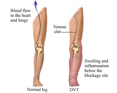
The first step when DVT is suspected is to obtain a venous duplex study. First line of therapy is of course anticoagulation in proper individual patient.  When suspecting ileofem DVT based on duplex, I would prefer immediate CT to further evaluate possible venacaval or ileofem DVT.

Vein Thrombosis

Treatment option at this point may include thrombolysis therapy.  For the past 20 years, thrombolysis therapy has been available; however, there is no solid evidence supporting this for acute DVT despite lots of successful case reports and non-official data.

In my opinion, catheter based thrombolytic therapy is worth trying in venacaval or ileofem DVT patients especially if they are young or good candidates.  There are several possible benefits.  One is to prevent post-thrombotic syndrome which may occur in 50% of patients.  Secondly it may prevent recurrent thrombosis.  Thirdly it can treat or identify underlying cause such as May-Thurner syndrome which is usually left iliac vein stenosis caused by right side common iliac artery compression.    Venogram can identify this defect after thrombolysis and it is best treated by stent application.

Thrombolysis therapy is time consuming procedure, however, usually taking 24 to 48 hours duration as well as requiring temporary IVC filter placement.  Bleeding complication is the most common down side of this procedure.  Again patient selection should be done carefully and full informations should be discussed with patients regarding detailed data of benefits and risks.

Carotid Stenting vs Surgery

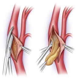
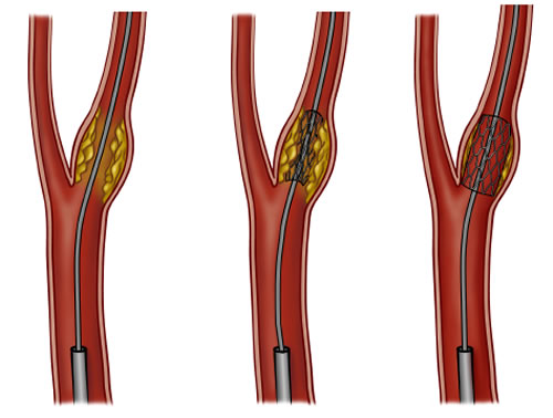
Recently, with a very well conducted, randomized study called CREST (Carotid Endarterctomy vs. Stenting Trial); there are various concerns among vascular physicians. Based on the study, current guidelines for carotid disease may differ in the future. FDA guideline currently dictates carotid endarterectomy as a standard of care for this disease, but carotid stenting may also be considered equally effective therapy in the future. There are several concerns about the CREST results, however. One of the interesting factors was the patient’s age. According to the report, patients aged 65 or younger did better with carotid stenting and older populations did better with open surgery. This is a difficult result to believe but was predicted; prior to full reports on CREST outcomes, preliminary reports identified the octogenarians group (those greater than 80 years-old), as doing poorly with stenting. Another issue with the CREST findings highlighted was the complication rates. Complication rates were similar with both carotid surgery and carotid stenting. The differences being that with stenting, the risk of stroke was much higher, while with open surgery, the risk of heart attack was much higher. Therefore, both groups exhibited similar post procedure co-morbid conditions and complications.

Indications for surgery may differ from those for carotid stenting. Based on NASCET (North-America Symptomatic Carotid Endarterectomy Trial), 70 % or more symptomatic patients would benefit from surgery. For asymptomatic patients, I would use the same guideline of 70 % or more  stenosis to treat surgically. Though, based on the ACAS (Asymptomatic Carotid Atherosclerosis Study), and the ACST (Asymptomatic Carotid Surgery Trial), asymptomatic patients with 60 % or more stenosis would benefit from surgery.

Despite all the above criteria and statistical facts, most surgeons will only operate at 80 % or greater stenotic disease.  However, morphology of the plaque and underlying atherosclerotic unstable plaque may need earlier intervention. Therefore, we would like to take care of disease at 70 % or higher since risks involved with carotid endartectomy are low. In my opinion, carotid endartectomy should still be the standard of care for carotid disease patients. With my experience, both stroke and heart attack events are extremely low after surgery.  Cardiac evaluation and clearance should always be done prior to surgery, as I do with my patients. This certainly minimizes chances of any major cardiac event.

In conclusion, I would like to convince primary care physicians and other specialists to emphasize the awareness of carotid disease. Again stroke rate for any stenosis greater than 70 % is significant based on all the above studies mentioned. There is a clear benefit with carotid surgery or stenting to prevent stroke. I usually use both duplex studies and angiographic feature to use in determining when surgery is indicated. Duplex criteria may differ from one to another, but I would like to use velocity and ratio criteria combination to select patients who fall into severe stenosis, generally greater than 70-80 %. Angiogram is almost always indicated to further assess the anatomical feature as well as to confirm the degree of disease or stenosis. As I always do, it is important to review films yourself to ensure an accurate reading.

Carotid disease treatment modality may change significantly in the near future. I believe that there is a vital role that carotid stenting can offer. Total Vascular Surgery would like to provide the best options in treatment for carotid disease, including open surgery and stenting in the near future. However, at the present time, only selective patients may be candidates for carotid stenting.

PAD (Peripheral Arterial Disease)

Peripheral Arterial Disease; previously known as ‘peripheral vascular disease’, refers mainly to lower extremity occlusive disease. The symptoms of this disease include; claudicating pain, rest pain, and/or non-healing wounds or sores.

Options for treatment of PAD traditionally include conservative medical management (such as walking exercise, cessation of smoking and lifestyle changes) or open surgery such as bypass or endarterectomy. However, with the recent advances in the endovascular field, there are many less invasive treatments available. These treatments include angioplasty, stenting, and artrectomy (excisional or laser). These options have recently been the first choice of treatment for PAD.

It is certainly true that endovascular treatment has replaced the traditional ways of treating the vast majority of patients. Mild to moderate claudicating symptoms in the legs can be well treated with walking regimen therapy, modifying life style; such as quitting smoking, eating a more healthy diet, control blood sugars if diabetic, control blood pressure, and avoiding stress. However, large number of patients with a focal lesion found on duplex studies may be a good candidate for simple angioplasty treatment the benefits of which can often last 2-3 years.

Indications for interventions in PAD includes disabling claudicating pain on walking generally these can be different from individual to individual but generally less than a block walking distance constitutes severe degree. Open sore or wound that is not healing due to PAD is another obvious indication. Rest pain is rare but another devastating indicator of critical PAD. Besides these indications, clinical judgment is often required to treat some advanced problems such diabetes induced critical PAD. These patients may not experience symptoms until they develop obvious signs of ischemia. Therefore, early referral can be a good tool to monitor this subset of patients.

Follow up with a vascular specialist after any kind of procedure is extremely important because stents, angioplasty, and bypass grafts do not last forever. This is a proven fact throughout numerous clinical studies and literature supports this idea as well. Therefore, procedures without follow up for many years can be very harmful for patients. In addition, recovery for re-do procedures in patients with stenosis or late recurrent disease are very difficult and often not possible.

Patients with mild to moderate claudicating pain should be referred to a vascular specialist for evaluation, monitoring, and treatment of this disease. When left untreated, PAD can escalate, causing severe complications. When caught early, conservative management and lifestyle changes (such as quitting smoking) can go a long way towards helping patients avoid more aggressive treatment. As always, follow-up after any procedure is the number one key to vascular health.

Dr. Lim accepts both inpatient and outpatient referrals.
For an Urgent Referral, please call 916-784-1836 and
we will get you in touch with Dr. Lim.
All other referrals; fax to 916-784-1880.
We have extended Dr. Lim’s availability in the Roseville Area to accommodate the growing need for a vascular surgeon.
As always thank you for your referrals.

Abdominal Aortic Aneurysm


Abdominal aortic aneurysm is one of leading causes of sudden death in the US. According to recent medical survey, despite modern technology and advanced medical and surgical treatment, death rate from ruptured abdominal aortic aneurysm is still very high at 40-50% among those who made to the surgery and 90% mortality rate is true for those including not being able to make it on time. Therefore, early detection and surgical treatment is essential in preventing death.

Most simple clinical diagnostic tool for abdominal aortic aneurysm detection is deep palpation at mid to left upper quadrant area. More than 90% of the time, a pulsatile mass should be appreciated in decent size aneurysms. This is a simple but very important clinical assessment skill for early detection of this deadly disease.

Of course, abdominal ultrasound screening tool is as equally important as clinical skill. Again vascular screening for early detection of abdominal aortic aneurysm is currently under study and the commonly known ‘Lifeline’ screening is one of them.

Etiologies for abdominal aortic aneurysm are multiple but atherosclerosis is the number one cause of this disease. Smoking, hypertension and hypercholesterolemia are the trio of enemy for atherosclerosis. Three most important concurrent medical conditions with AAA are coronary artery disease, Chronic Obstructive Pulmonary Disease and hypertension. Therefore, it is important to control hypertension and underlying lung conditions if anyone is diagnosed with AAA and in order to prevent worsening expansion. 15% of time it can be inherited as well. Cardiac evaluation is a must even if there is no history of cardiac disease in the family.

Rupture rate statistically rises suddenly beyond 5cm. However, this is very controversial because every one has different ‘normal size’ of aorta. Generally, in female patients, 5cm would be much too big compared to same size in male patients. However, 5cm is universally accepted size for timing of intervention. In my practice, I tend to individualize the timing for aneurysm repair; in other words, sizing is not the only criteria for intervention. Medically high risk male patients can be postponed until they reach 5.5cm. However, size is not the only indication for aneurysm repair. The next most common indication for AAA repair is rapid expansion. Generally, an AAA grows 4mm per year. If there is an expansion faster than 4mm per year, my recommendation would be early intervention. Close observation and follow up is very important for AAA patients. Ultrasound or duplex vascular examination is very important not only for clinicians but also for patients. There can be up to 5mm difference in measurement depending on technicians’ skill and experience.)

Other indication for AAA repair includes heavy ulcerated plaque protruding out asymmetrically in the aneurysm. We call this “saccular aneurysm.” Usual aneurysm shape should be ‘fusiform’ which means uniform and symmetrical dilatation of aneurysm. Saccular aneurysm is non-symmetrical, meaning that one portion of aneurysm could be excessively protruding out and this can be very dangerous even if the size is not quiet 5cm in maximum diameter. This portion of aneurysm can rupture easily due to that fact that is so inflated.
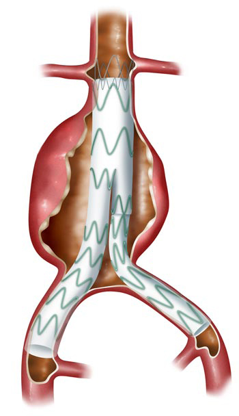
Aortic2Symptomatic aneurysm should be repaired regardless of size but it is very unusual to have symptoms such as abdominal or back pain in small size aneurysm. Some female AAA patients with small body habitus should be recommended for early aneurysm repair probably even at 4.5cm.

There are other indications for AAA repairs but the most aneurysm patients should be referred to a qualified vascular specialist who can treat AAA and follow-up after such treatment.

Iliac aneurysm is often found in AAA patients at the same time, in most cases. Iliac artery aneurysm can rupture as well if not treated at right time. 2.5 – 3cm in size is currently acceptable for timing of repair. Therefore, if a patient has 4cm AAA and 3cm iliac aneurysm in one side, this patient will require surgery. Common iliac artery aneurysm is the most common aneurysm followed by hypogastic or internal iliac artery aneurysm. If ultrasound study is not specific about iliac portion of study, one should note that there is a 50-70% chance of concurrent aneurysm at iliac arteries along with AAA.

Surgical treatment for AAA is traditional open repair vs. endovascular repair. 80% of time or even higher with recent advancement of technology, endovascular repair is very possible. Short neck or angulated neck and even with 15% or more thrombosed neck could be accepted for endovascular repair with newly available devices. More and more non-invasive options are available today than 3-5 years ago. Of course, a close follow up is very important because of possible complications with endovascular stentgraft repair. Endoleak is the most common complication after endovascular repair, occurring in up to 10% of patients. This may require years of follow up and sometimes those individuals with persistent endoleak may require additional treatment.

In summary, early detection can be easily performed by placing your hand in patient’s abdomen specifically looking for pulsatile mass. In normal individual, it should not be easily felt on deep palpation. Next, ultrasound study is very important tool to determine the size. Referral to an appropriately qualified vascular surgeon is very important for not only treatment but follow up as well.

Dr. Lim accepts both inpatient and outpatient referrals.
For an Urgent Referral, please call 916-784-1836
and we will get you in touch with Dr. Lim.

All other referrals; fax to 916-784-1880.
We have extended Dr. Lim’s availability in the Roseville Area to accommodate the growing need for a vascular surgeon.

As always thank you for your referrals.
Total Vein Care

Venous disease is the most common vascular problem in the nation. Among them, varicose vein problem is the most common diagnosis in most Americans both male and female. According to recent statistics, at least 40-50% of American females older than age 45 suffer from some degree of varicose vein problem. With recent developments of the endo-venous techniques, varicose vein treatment has become easier than ever. Office based varicose vein practices have been amongst the most the exciting fields of development for qualified physicians. However, true understanding and knowledge based venous disease diagnosis and their appropriate treatments are very important.

Clinical symptoms of venous disease can be very broad. Heaviness or early fatigue on standing can be symptoms of venous insufficiency. Among patients diagnosed with Restless Leg Syndrome, most have been identified as having venous disease as well. Other signs and symptoms of venous disease include swelling, chronic leg pain without clear etiology, cellulites, ulceration, bruises, skin discoloration and so on. Obvious varicose vein protrusion is not the only condition you should be concerned about. Some patients may have swelling problem without any obvious varicosities. However, their superficial vein system can be refluxing and they could be a good candidate for surgical or endovascular treatment.

Radiofrequency ablation therapy has been accepted as one of most popular and credited treatments available today. I personally believe that radiofrequency ablation therapy is a better choice than laser therapy. Greater and lesser saphenous veins, anterior lateral thigh accessory branch and perforators can be treated using this technique. These therapies can be performed in the office setting with safe technique and experience. Individual varicosities striping or phlebectomies can be performed as well at office setting. Of course, individual evaluation and recommendation is very important. Patients with excessive varicosities and low tolerance to pain should be recommended to do phlebectomies in the hospital. Sclerotherapy is another very common treatment which can be offered to patients. Each individual may require 3-6 sessions of treatment depending on the severity and recurrence. Unfortunately sclerotherapy for spider vein or telengiectasia has been categorized as cosmetic treatment and therefore is not reimbursed from insurance companies.

Overall, vein care should be provided to each individual because venous disease can be a life time problem and simple office based treatments can change a patient’s quality of life.

Total Vascular Surgery is going to start a ‘total vein care clinic’ in the year 2009. We have a dedicated vascular lab with a qualified and experienced technician especially for vein disease. We will do our best to provide the total vein care for all your patients. Please make a referral to Dr. Lim if you have patients with chronic leg pain, swelling, varicose vein problem, discoloration, ulceration, fatigue, discomfort, unexplained symptoms, non-healing wounds, phlebitis, restless leg syndrome…etc.